Tunnel view (Holmblad method, Beclere method, Camp coventry method)
Holmblad method
Beclere method
Camp coventry method
Holmblad method
Purpose
This method can evaluate the femoral intercondylar fossa, femoral condyle, tibial epiphyseal joint surface, bone and cartilage lesions, and narrowing of the joint space.
The Holmblad method is designed to view intra-articular free bodies that cannot be observed in the frontal or lateral views.
Prior confirmation
Remove obstacles.
Positioning


On all fours posture.
The femoral axis is tilted forward 20° and the angle between the femoral axis and the bed is 70°.
A pillow is placed at the ankle, and the angle between the lower leg axis and the bed is 10°.
The femoral axis is tilted to account for the cervical body angle and the medial side of the intercondylar fossa is projected tangentially (p18).
A shielding apron should be placed over the buttocks to protect against gonadal exposure.
CR, distance, field size
CR : X-rays are directed vertically 1.5 cm below the inferior edge of the patella.
Distance : 100cm
Field size : The range fully includes the knee joint.
Exposure condition
58kV / 8mAs
Grid ( – )
Image, check-point
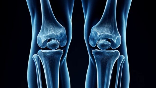
Normal (Fig.4)


The intercondylar fossa must be projected tangentially.
The patella should not overlap the intercondylar fossa.
There should be no rotation of the knee joint (symmetry of the medial and lateral femoral condyles and 1/2-1/3 of the fibular head overlapping the tibia).
Intercondylar ridge must be observable inside of intercondylar.
The patella should be observable overlapping the femur.
Observable tolerance of soft tissue and fat around the knee joint in addition to bony tissue.
Videos
Related materials
Holmblad Variations
Beclere method
Purpose
Image distortion due to oblique incidence.
Image quality deteriorates due to the distance between the subject and the cassette.
Increased gonadal exposure compared to other methods.
Thus, not a preferred imaging method however Useful for patients who can only lie on their back.
Extensive observation of the supra-tibial articular surface and the intercondylar fossa.
Excellent for observation of knee locking and intercondylar eminence due to free bodies.
Prior confirmation
Remove obstacles.
Positioning


Back supine position.
Flex the knee joint 40°.
Internal rotation so that the line connecting the medial and lateral femoral condyles is parallel to the cassette.
Position the cassette with oblique incidence in mind.
The cassette and the back of the knee should be as close as possible.
Use a shielding apron for gonadal protection.
CR, distance, field size
CR : Oblique incidence at 30°-40° in a caudal direction toward a point 1.5 cm from the inferior edge of the patella.
Distance : 100cm
Field size : The range shall include the knee joint completely.
Exposure condition
58kV / 8mAs
Gird ( – )
Image, check-point
Normal (radiopaedia)


The patella should not overlap the intercondylar fossa.
The medial and lateral condyles of the femur and tibia should be symmetrically drawn.
The plane of the knee joint should be centered.
The patella should overlap the femur and be visible.
Tolerance for soft tissue and fat around the knee joint in addition to bony tissue can be observable.
Videos
Related materials
Camp conventry method
Japanese ver.
Rad tech on duty
ce4rt
Purpose
The intercondylar fossa, the posterior inferior aspect of the femoral condyle, the medial and lateral intercondylar tuberosities, the intercondylar ridge, and the knee joint space are visible. Evaluation of intra-articular free bodies and cartilage damage.
Prior confirmation
Remove obstacles.
Positioning


Prone.
The knee on the examining side is flexed 40° and placed on a support table to be held.
Position the cassette so that the knee joint plane is projected centrally, taking into account oblique incidence in the cephalocaudal direction.
The knee joint is internally rotated so that the knee joint is in mid-position.
CR, distance, field size
CR : Oblique incidence at 40° in a caudal direction, passing 1.5 cm below the inferior edge of the patella.
Distance : 100cm
Field size : Include the knee joint completely.
Exposure condition
58kV / 8mAs
Grid ( – )
Image, check-point
Normal


The intercondylar fossa should be widely delineated.
That there is no rotation of the knee joint (symmetry of the medial and lateral femoral condyles and 1/2-1/3 of the fibular head overlapping the tibia)
The patella should not overlap the intercondylar fossa.
Intercondylar ridge should be observable.
The patella should overlap the femur and be observable.
Observable tolerance of soft tissue and fat around the knee joint in addition to bony tissue.
Videos
Related materials
Various X-ray views of Knee Joint (p36-45)