Thoracic spine AP view
Purpose
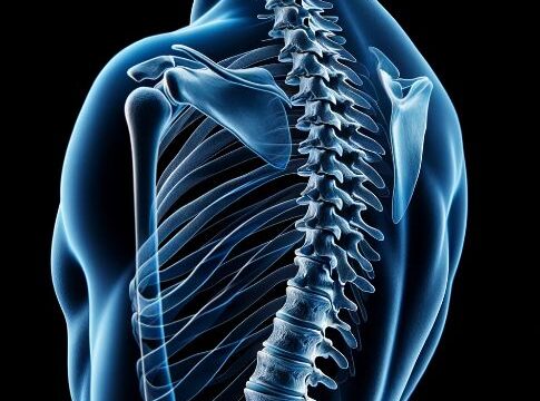
Observation of intervertebral disc, thoracic vertebral bodies, posterior ribs, and costovertebral joints.
Observation of compression fractures, subluxations, and kyphosis.
Prior confirmation
Confirm whether the patient is to be radiographed in the standing or supine position.
Remove any obstacles.
Positioning


Erect :
Standing position in AP orientation, with lower extremities shoulder-width apart.
The coronal plane and the image receiving plane should be parallel to each other to avoid body twisting.
Align the mid-sagittal plane with the central axis of the cassette.
The cassette should be 14×17 inchs size and aligned with the midpoint of the suprasternal fossa-xiphoid process (subscapular angle) in order to achieve a center height of Th7.
The upper end of the cassette should be aligned under the thyroid cartilage ridge.
Supine :
Supine position. Chin up without pillow.
Keep the coronal plane parallel to the image-receiving surface to avoid twisting of the body.
Align the mid-sagittal plane with the central axis of the cassette.
Bend both knees to reduce kyphosis of the thoracic spine.
Use a 14×17 inchs size cassette and align the cassette with the midpoint of the suprasternal fossa and the xiphoid process (subscapular angle) in order to set the center height at Th7.
The upper end of the cassette should be aligned under the thyroid cartilage ridge.
CR, distance, field size
CR : Vertical incidence at the midpoint of the suprasternal fossa – xiphoid process.
Distance : 100-150cm
Field size : The top and bottom sides should be widened to 14×17 inchs size. The left and right sides should be minimized according to the purpose of imaging.
The upper thoracic spine is overexposed compared to the lower thoracic spine, so the heel effect should be used (anode toward the head) or a wedge filter should be used.
Exposure condition
80kV / 10mAs
(Using high voltage reduces contrast and increases the range of observation of the upper and lower thoracic spine.)
Grid ( + )
Suspend respiration on expiration.
Image, check-point
Normal (Radiopaedia)


From C7 to L1 must be included.
The spinal column should be projected in the center of the image/irradiation field.
The vertebral limb, intervertebral space, vertebral arch roots, spinous processes, transverse processes, and posterior ribs should be observable from top to bottom.
The vertebral arch roots should be symmetrical, the spinous processes should pass through the center of the vertebral body, and there should be no torsion in the body axis.
Videos
Related materials