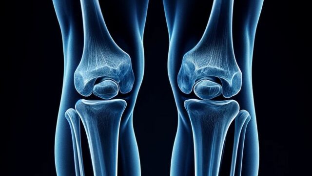
Knee joint oblique view (Internal/external rotate)
Internal rotate
External rotate
Internal rotate
Purpose
Oblique view is used for close examination of tibial plateau fractures and proximal tibiofibular joints.
It is used as an additional imaging because it can reveal diseases that cannot be seen on the AP or LAT views.
It is useful in facilities where CT scans are not available.
It is particularly suitable for observation of the anterior medial and posterior lateral aspects of the knee joint.
For oblique imaging in only one direction, the internal rotation position is selected.
Prior confirmation
Remove obstacles.
Positioning


Supine position.
Extend the knee joint.
Rotate not only the knee but also the entire trunk and lower leg so that the entire lower leg is in an oblique position.
Internal rotation so that the line connecting the medial and lateral femoral condyles is 45 degrees.
CR, distance, field size
CR : Vertical incidence 1.5 cm below the inferior edge of the patella and toward the lateral side of the patella.
Distance : 100cm
Field size : The range includes the distal 1/3 of the femur to the proximal 1/3 of the lower leg, narrowing to the skin surface on the left and right sides.
Exposure condition
55kV / 5mAs
Grid ( – )
Image, check-point
Normal


The tibia and lateral femur are widely observed without overlapping the patella.
Half of the patella protrudes away from the femur.
The tibiofibular joint can be observed without the fibula overlapping other bones.
In addition to the bony tissues and bony beams, soft tissues and fat around the knee joint can be observed.
Videos
Related materials
External rotate
Purpose
It can be used as an additional imaging to detect diseases that cannot be seen on the AP and LAT views.
Useful in facilities where CT scans are not available.
Particularly excellent for observation of the medial aspect of the knee joint.
Prior confirmation
Remove obstacles.
Positioning


Supine position.
Extend the knee joint.
Rotate not only the knee but also the entire trunk and lower leg so that the entire lower leg is in an oblique position.
External rotation so that the line connecting the medial and lateral femoral condyles is 45 degrees.
CR, distance, field size
CR : Vertical incidence 1.5 cm below the inferior edge of the patella and toward the medial side of the patella.
Distance : 100cm
Field size : The range includes the distal 1/3 of the femur to the proximal 1/3 of the lower leg, narrowing to the skin surface on the left and right sides.
Exposure condition
55kV / 5mAs
Grid ( – )
Image, check-point
Normal


The tibia and medial femur are widely observed without overlapping the patella.
About half of the patella is projected away from the femur.
The fibula is observed completely overlapping the tibia.
Tolerance for soft tissue and fat around the knee joint can be observed in addition to the bony tissue and bony trabeculae.
Videos
Related materials