Inlet view, Pubic axial view
Purpose
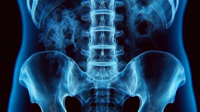
Observe the pelvis ring in an front view.
The pubic axial view differs only in the CR and irradiation field.
Observe the anterior-posterior displacement of the pelvic ring due to trauma.
Prior confirmation
Confirm with a doctor if there is a suspicion of pelvic fracture and if it is safe to move the lower limbs.
Remove any obstacles.
Positioning


Supine position.
Align the mid-sagittal plane and the center axis of the cassette.
Extend both lower limbs (or slightly flex them).
To eliminate pelvic tilt, ensure that both left and right anterior superior iliac spines are equidistant from the cassette.
Rotate both knee joints 20° internally, only if there is no suspicion of fracture.
Place the upper limbs away from the irradiation field.
Position the cassette so that the exit point and center coincide.
Attach the R/L markers.
CR, distance, field size
CR :
For the inlet view, the crosshairs of the radiation field should pass through a point 3 finger-width cranial to the greater trochanter at an oblique angle of 30°-40° in the cranio-caudal direction.
For the pubic axial view, the crosshairs of the radiation field should pass through the greater trochanter at an oblique angle of 30°-40° in the cranio-caudal direction.
Distance : 100-130cm
Field size : Narrow the field to include the skin surface on both sides and the area from the iliac crest to the lesser trochanter. For the pubic axial view, the field size should be minimized to the necessary extent.
Exposure condition
75kV / 25mAs (or 90kV or higher for dose reduction).
Grid ( + ). *Pay attention to the direction of the grid foil for oblique incidence.
Suspend respiration.
Image, check-point
Normal (Radiopaedia)


Include the area from the iliac crest to the lesser trochanter.
Ensure that the iliac and ischial spines are projected symmetrically on both sides.
The pelvic cavity should be projected at the center of the radiation field.
The ilium should be clearly visible (without overexposure).
The pubis and ischium should overlap, and the obturator foramen is seldom observed.
Soft tissues should be visible.
There should be no blur.
Videos
Related materials