
Cervical spine open mouth view
Purpose
C1, C2, and soft tissues are observed.
Observations of Jefferson fracture and odontoid process fracture.
Prior confirmation
Always observe lateral images and obtain a physician’s permission before taking radiograph with the mouth open.
In case of trauma, avoid hyperextentional view.
If an open mouth is not possible, consider the Fuchs or Judd technique.
Remove any obstructive shadows.
(If hair is tied up, bundle it or remove necklaces, hairpins, etc.)
Positioning

Erect or supine.
The patient head should be viewed from the cephalic side to avoid head torsion, and the sagittal plane should be perpendicular to the imaging detector.
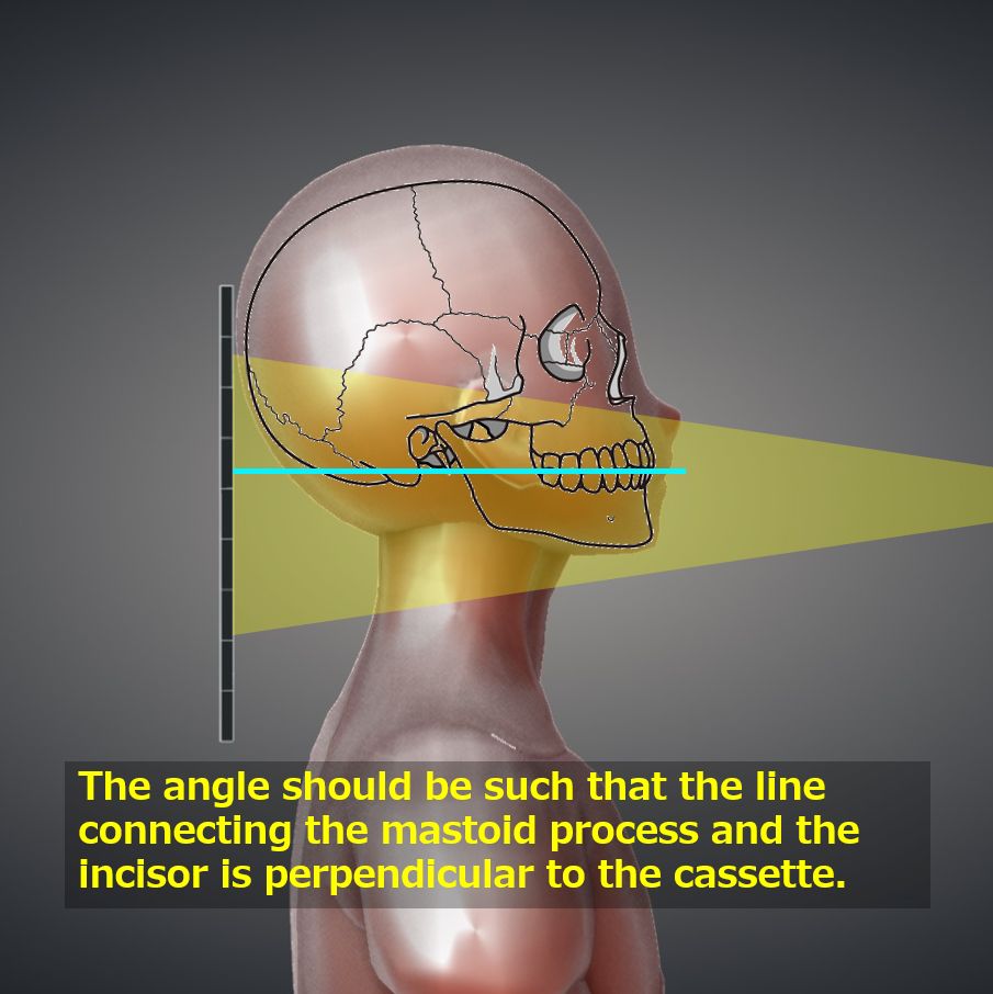
The vertical center of the cassette should be aligned with the height of the mastoid process.
At maximum aperture, the angle of head should be such that the line connecting the inferior margin of the maxillary incisors with the mastoid process (or the line connecting the nasal root with the external auditory meatus) is perpendicular to the imaging detector. If this is not possible, the angle of incidence should be adjusted.
To minimize the time between opening the mouth and exposure, set the imaging conditions in advance.
The tongue should be lowered toward the mandible to avoid obstructive shadows.
CR, distance, field size
CR : Vertical incidence toward the center of the open mouth.
Distance : Nearest-100 cm.
Field size : The head side should be narrowed so as not to include the eyes. The left and right sides should be extended to the skin surface.
Since the open mouth often moves, observe the patient carefully and press the exposure switch when the patient is not moving.
Exposure condition
55kV / 5mAs
Grid ( – )
Suspend respiration.
Image, check-point
Normal (Radiopaedia)
Jefferson fracture (Radiopaedia)


The lateral mass and transverse process (C1) should be observable.
The odontoid process (C2) should not be overexposed and the vertebral body should be observable.
The gap of the atlantoaxial joint should be symmetrical and wide.
The upper incisors and occipital bone should not overlap C1/C2.
Since overlap of the obstruction to the target area may be unavoidable depending on the skeletal structure, other imaging methods (Fuchs or Judd method) or CT should be considered.
Videos
Related materials
Close mouth view
Fracture Classification : Anderson’s Classification
(Type 1) Oblique fracture of the tip of the dental process
(Type 2) Fracture up to the base of the odontoid process
(Type 3) Fracture extending to the C2 vertebral body
Type 2 is prone to pseudoarthrosis and requires surgical intervention.


