Purpose
In a true AP view, the joint space on the fibular side is not as clearly visualized. In contrast, this imaging technique (mortise view) allows for a wider depiction of the joint space. In many countries, the mortise view is taken as the AP view.
This view is used to observe the distal tibia, distal fibula, ankle joint, and the base of the fifth metatarsal.
It is employed for the evaluation of fractures, dislocations, inflammations, and bone tumors.
Prior confirmation
Remove any obstacles.
If there is pain, do not force internal rotation or dorsiflexion of the lower limb.
Positioning
Sitting or supine position.
Extend the lower limb on the examined side and abduct the non-examined side to move it out of the irradiation field.
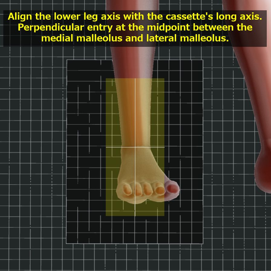
Align the lower leg axis on the examined side with the cassette’s long axis.
Align the ankle joint with the center of the cassette.
Dorsiflex the foot to make the sole of the foot and the cassette perpendicular.
Internally rotate the knee joint by 10° to 15°.
Make the line connecting the calcaneus and the fourth toe perpendicular to the cassette.
Place the RL marker.
CR, distance, field size
CR : Perpendicular to the midpoint of the line connecting the medial malleolus and lateral malleolus.
Distance : 100cm
Field size : Include the upper and lower regions from the distal one-third of the lower leg to the base of the fifth metatarsal. Narrow the field to the skin surface on the left and right sides.
Exposure condition
50kV / 4mAs
Grid ( – )
Image, check-point
Normal (Radiopaedia)
Ensure clear visualization of the talar dome (where all three sides measure 3-4mm, with over 2mm enlargement being abnormal).
Observe the distal tibiofibular joint space (normal if 6mm or less).
Clearly visualize the cortex and trabecular bone.
Include the distal tibiofibular joint and the base of the fifth metatarsal.
Ensure no overlap of the non-examined lower limb.
Maintain sufficient tolerance for soft tissue visualization.
Ensure the presence of the RL marker.
Prevent motion-induced blur.
Videos
Related materials
Weber’s fracture classification (used to determine treatment):
A: Distal to the syndesmosis of the tibia and fibula.
B: At the level of the syndesmosis of the tibia and fibula.
C: Proximal to the syndesmosis of the tibia and fibula.


