Purpose
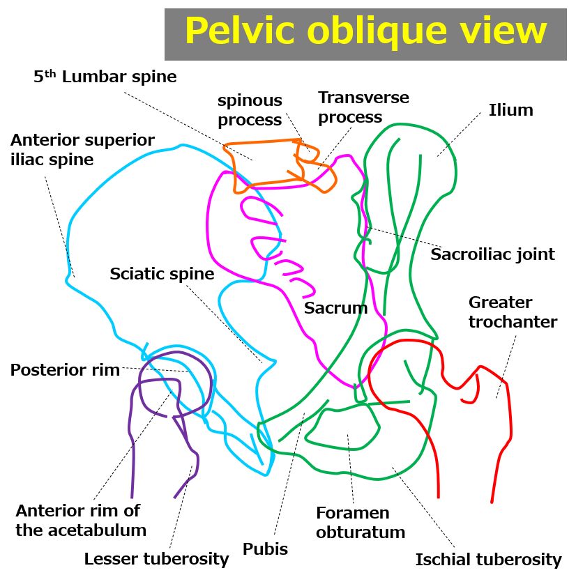
Observation of the acetabulum and ilium is performed. Depending on the radiation field and X-ray center, it is called that pelvic oblique view, ilium AP view, or ilium axial view.
Prior confirmation
Please confirm which method is ordered from the following: 1. Pelvic oblique view, 2. Ilium AP view, or 3. Ilium axial view.
Remove any obstructing objects.
Positioning
1. Pelvic oblique view : The patient lies supine with the non-affected side elevated 45 degrees.
2. Ilium AP view : The patient lies supine with the non-affected side elevated 45 degrees.
3. Ilium axial view : The patient lies supine with the affected side elevated 45 degrees.
The hip and knee joints on the side closer to the cassette are slightly flexed.
The leg on the side farther away from the cassette is flexed and abducted.
Align the incident point with the center of the cassette.
CR, distance, field size
CR :
1. Pelvic oblique view : At the midpoint between the left and right sides of the pelvis, perpendicular to a point 3 fingerbreadths above the greater trochanter.
2. Ilium AP view : Perpendicular to a point 2 cm posterior and 5 cm medial to the upper anterior iliac spine on the affected side.
3. Ilium axial view : Perpendicular to a point 2 cm posterior to the upper anterior iliac spine on the affected side.
Distance : 100-130 cm
Field size :
1. Pelvic oblique view : Includes both sides of the pelvis.
2. Ilium AP view : Includes the affected side’s ilium, ischium, and pubis.
3. Ilium axial view : Includes the affected side’s ilium, ischium, and pubis.
Exposure condition
80kV / 25mAs (or 90kV or higher for dose reduction)
Grid ( + )
Suspend respiratory
Image, check-point
Normal (Radiopaedia)
Ensure that the anterior and posterior rims of the acetabulum are visible.
Ensure that the thigh on the side farther from the cassette is not overlapping.
If both sides are being imaged, ensure that they are symmetrically captured.
The obturator foramen on the side closer to the cassette is not visible, while the obturator foramen on the farther side should be clearly visible.
The joint space between the acetabulum and femoral head should be uniform.
The iliac crest and upper anterior iliac spine should be visible without overexposure.
Soft tissues should be visible.
There should be no motion blur.
Videos
Related materials


