
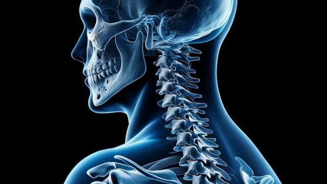
Cervical spine hyperflexion and hyperextension view
Purpose
Exclusion of whiplash and follow-up after spinal fusion surgery.
Observation of rheumatoid arthritis.
Confirmation of neck movement for insertion of endotracheal tube.
Observation of vertebral body range of motion, displacement of the vertebral arch, narrowing and enlargement of the vertebral canal space, dislocation and subluxation.
Prior confirmation
Follow the doctor’s instructions, as bending the neck forward or backward can cause neck injury.
Remove any obstalces.
Positioning


Sitting or rect position. Stabilize the posture with the sides to the cassette.
Keep the sagittal plane parallel to the cassette so that there is no twisting of the body.
Check from the front that the neck (and body) is not bent.
Adjust the height of the cassette so that the 4th C-spine (laryngeal ridge) is centered.
Grasp the chair with both hands and lower both shoulders.
Hyperflexion : Bring the chin as close to the chest as possible, without tipping the back forward.
hyperextension : Bring the occipital bone as close to the back as possible with the chin up, without tipping the back.
CR, distance, field size
CR : Horizontal incidence at the level of the 4th cervical vertebra (laryngeal ridge) toward the anterior-posterior center of the neck.
Distance : 100-150 cm.
Field size : The upper and lower areas should include the external auditory foramen to the spinous process of the seventh cervical vertebra. The left and right sides should be extended to the skin surface.
Since the patient often moves, observe the patient carefully and press the exposure switch when the patient is not moving. The imaging conditions should be set in advance.
Exposure condition
74kV / 16mAs
Grid ( + )
Suspend respiration on full expiration.
Image, check-point
Normal (Radiopaedia)
Normal (wikimedia)


The image should not be blurred.
The external auditory foramen to the 1st thoracic vertebra (2nd thoracic vertebra if possible) should be observable.
The fourth cervical vertebra should be centered in height.
Laterality of the neck should be maintained without twisting. (Judged by the superior and inferior articular processes of the vertebral body and the vertebral body and mandibular angle)
A wide intervertebral space can be observed.
The right and left intervertebral joints should overlap.
The spinous processes can be observed laterally.
Wide spacing of spinous processes can be observed in anterior flexion, and narrow spacing of spinous processes can be observed in posterior flexion.
Soft tissues should be observable.
Videos
Related materials


