Calcaneus axial view
Purpose
Observation of the calcaneus, posterior talocalcaneal joint, and middle talocalcaneal joint.
Assessment for fractures, dislocations, inflammation, and bone tumors.
Prior confirmation
Remove any obstacles.
Do not force dorsiflexion if there is pain.
Positioning


Supine position.
Extend the lower limb on the examined side.
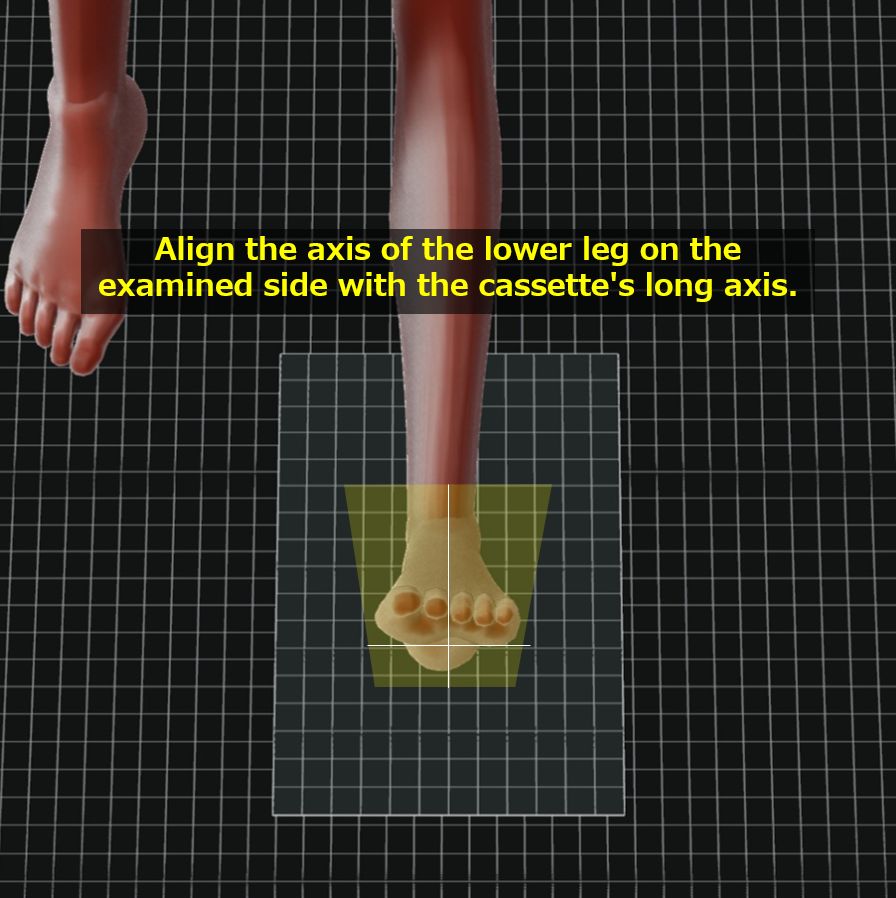
Align the axis of the lower leg on the examined side with the cassette’s long axis.
Ensure that the foot reference line (a line connecting the calcaneus and the second toe) is perpendicular to the cassette.
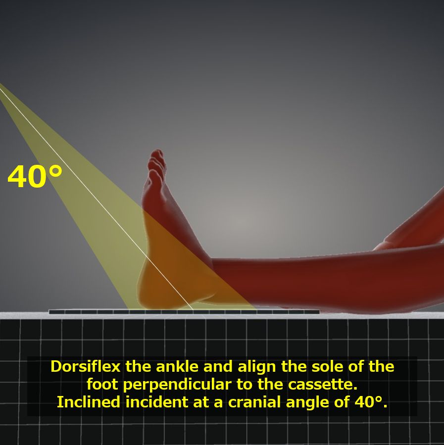
Dorsiflex the foot on the examined side, bringing the sole of the foot to a 90° angle (or maximum dorsiflexion) relative to the lower leg axis. Use a towel or similar if necessary to assist with the movement.
If there is pain or inability to dorsiflex:
1. Increase the incidence angle.
2. Tilt the axis of the lower leg.
Align the center of the calcaneus with the cassette center by observing the shadow within the irradiation field.
Place the R/L marker.
CR, distance, field size
CR : Inclined incident at a cranial angle of 40 degrees toward the base of the third metatarsal.
Distance : 100 cm
Field size : Including the calcaneus and metatarsal bones.
Exposure condition
58kV / 10mAs
Grid ( – )
Image, check-point
Normal (Radiopaedia)


The posterior talocalcaneal joint and middle talocalcaneal joint are clearly observable.
The calcaneal tuberosity is not overexposed.
Cortical and trabecular bone structures are clearly visible.
The base of the fifth metatarsal is included.
There is a tolerance for observing soft tissue.
An R/L marker is present.
There is no motion-induced blur.
Videos
Related materials