Purpose
Alternative AP view in patients can not extend the elbow.
Prior confirmation
Confirm whether the target part is humerus or forearm.
Positioning
Seat the patient in the chair.
Raise the upper arm and elbow joint to shoulder height.
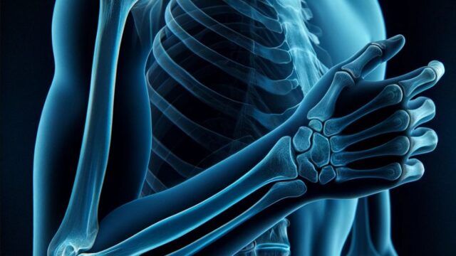
The elbow joint is in maximum flexion and the fingers touch the shoulder.
The back of the upper arm should adhere to the film.
Overlap the forearm and upper arm axis.
The medial and lateral epicondyles should be equidistant from the film.
CR, distance, field size
CR :
1. Observe the humerus : Incident perpendicular to the film, midway between the medial and lateral epicondyles.
2. Observe the forearm : Incident perpendicular to the forearm axis at a point 5 cm distal to the olecranon.
Distance : 100cm
Field size : Irradiation field includes the distal 1/3 of the humerus to the proximal 1/3 of the forearm, narrowing to the skin surface on the left and right sides.
Exposure condition
54kV / 5mAs
Grid ( – )
Image, check-point
1. Observe the humerus
2. Observe the forearm
Videos
Related materials


