
Chest lateral view
Purpose
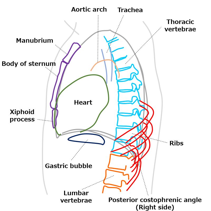
Observation of the trachea, aorta, heart, sternum, lung fields, diaphragm, and posterior costophrenic angle.
Depending on the purpose of the examination, it is usually imaged in the RL direction to minimize heart enlargement.
Prior confirmation
Check for the presence of scoliosis from the frontal PA image.
Remove any obstructing objects (such as necklaces).
Positioning


Upright stance with the left side in close contact with the cassette.
Ensure that the coronal plane is perpendicular to the cassette.
The height of the cassette should be the same as for the frontal PA view (upper edge 5cm above the shoulder joint).
Place markers (R->L, L->R) on the cassette.
Raise both arms sufficiently to avoid obstructing the upper lobes of the lungs.
Slightly tilt forward and raise the chin.
CR, distance, field size
CR : The X-ray should be incident to the cassette perpendicularly. The center of the X-ray beam should be positioned at the midpoint of the trunk and at the height of the scapular angle during relaxation.
Distance : 200cm.
Field size : Centered on the scapula angle, extending to the lower border of the rib arch (having a rough idea of the approximate location of the posterior costophrenic angle from the frontal PA view).
Exposure condition
120kV / 8mAs. (To minimize the influence of cardiac motion, the exposure time should be kept as short as possible (maximizing the mA)).
Grid ( + )
Maximum inspiration
Image, check-point
Normal (Radiopaedia)

Ensure that there are no areas of missing or obscured lung fields in the image.
Ensure that both arms and the chin are not overlapping with other structures.
Check if the vertebral bodies are projected with tangents.
Observe if the hilar region, which overlaps with the heart, is visible.
If taken at maximum inspiration, ten posterior ribs should be visible above the diaphragm (alternatively, compare the diaphragm position with the frontal PA image).
The posterior ribs on the side farther from the cassette and the rib costophrenic angle may appear slightly enlarged and projected posteriorly.
Ensure there is no blurring present.
Videos
Related materials


