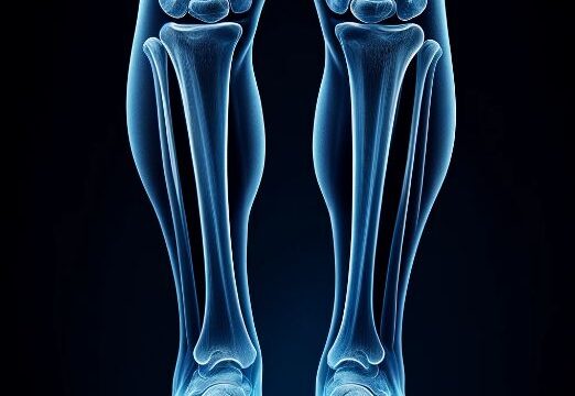
Lower leg AP view
Purpose
Projecting from the knee joint to the ankle joint and observing the tibia and fibula from the front.
Diagnosing dislocation, fracture, osteomyelitis, and foreign bodies.
Prior confirmation
Remove any obstacles.
Positioning


Supine position.
No pelvic tilt and equal distance between the bilateral anterior superior iliac spines and patient table.
Internally rotate the lower limb to orient the joint (closer to the affected area) towards the front.
Knee Joint : Align the line connecting the medial and lateral condyles of the femur parallel to the cassette.
Ankle Joint : Align the line connecting the medial malleolus and lateral malleolus of the tibia parallel to the cassette.
Place the lower leg parallel to the long axis of the cassette with 17×14 inch size. If this is not feasible, position the lower leg diagonally.
Dorsiflex the ankle to achieve a 90° angle at the ankle joint.
Abduct the non-examined lower limb and keep it out of the field of radiation.
Considering the heel effect, orient the anode of the X-ray tube towards the ankle joint side.
Cover any unnecessary radiation field with a lead plate.
Position the RL marker.
CR, distance, field size
CR : At the midpoint between the knee joint and ankle joint, perpendicular to the skin surface on the fibular side.
Distance : 100cm
Field size : Include the knee joint to the ankle joint, narrowing down to the skin surface.
Exposure condition
55kV / 5mAs
Gird ( – )
Image, check-point
Normal (Radiopaedia)


Ensure that the area from the knee joint to the ankle joint is included.
The fibula should overlap with the tibia both proximally edge and distally edge, but there should be no other overlapping.
Cortical and medullary areas of the bone shaft should be clearly visible.
The non-examined lower limb should not overlap.
There should be sufficient tolerance to observe soft tissue.
The RL marker should be visible.
There should be no motion-induced blur.
Videos
Related materials